Video Details
|
The Magnetic Mallet and its Use in Daily Practice - Clinical Perspective
Description:
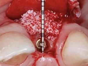
In our implant daily practice we often have to deal with conservative root’s extractions, thin crestal bone and poor quality alveolar bone, especially in the posterior areas where it is often necessary to increase the height of the residual bone after dental loss. We know many techniques to manage those situations, like guided bone regeneration with resorbable and non resorbable membranes, block bone grafts, cortical lamina with autogenous bone, crestal and lateral sinus lift, horizontal and vertical split ridge techniques. To perform the split crest technique and the crestal sinus lift we have used during the years many kind of osteotomes and bone expanders with manual hammers with great results in terms of quality and quantity amount of bone achieved with these techniques, and especially better morbidity during the healing period and the necessary costs in time and money for the patients. The aim of this lecture is to analyze the use of an osteotome technique that I perform using a machine that is more precise than the traditional manual mallet, that you can manage using both hands. The magnetic mallet has several tips of different shapes and diameters to manage the native bone and to move it apically and around the implant.
The Magnet Mallet is a device based on a EU/US patent of invention in oral surgery. It uses magnetism to generate a very important acceleration of the osteotomes that maximizes the forces applied to the bone (making the procedure fast and efficient) and in the meantime minimizes the inertia and discomfort for the patient. Such speed doesn’t cause any bone heating therefore irrigation water is not needed.
Date Added:
9/1/2021
Author(s):
Fabio Manuel Filannino, DDS
Dr. Fabio Manuel Filannino
Professional Experience:
- 1998: Dentist at Dental Department and the Italian Navy - Lo...
[read more]
|

|
Online Videos / Surgery / Other Surgical / The Magnetic Mallet and its Use in Daily Practice - Clinical Perspective
Questions & Comments
|
Fabio Manuel Filannino - (9/1/2019 4:44 PM)
Thank you Dr Salama, I’m glad you found it interesting. The device has many potentials beside what already has in the daily practice. To answer to Haitham: a conical Implant and under preparation of the site is what we do to achieve primary stability and perform one stage surgery the most of the cases. So you decide following the insertion torque and the correct 3D position of the fixture according to the literature, based on what is the amount of the native bone you deal with. Thank you
|
Maurice Salama - (8/31/2019 2:26 PM)
Extremely Scientific and Clinically based presentation. I have recently had the opportunity to add this device into my clinical practice and have been impressed with the many opportunities to institute these concepts on a daily basis. Well done. Dr. Salama
|
Haitham Menim - (8/31/2019 9:09 AM)
how you u can be sure your implant will not be (sucked in)the sinus ?
When you decide to stop your implant inestion following magnatic mallet sinus elevation?
thanks
|
Related Videos |
|
|
Full Arch Immediate Loading Using Pterygoid Implants - Part 1 of 2
Non-grafting alternatives for the rehabilitation of the Atrophic Maxilla is a growing arena in our implant patient population.
The Intermaxillary Surgical Protocol for Zygomatic Implants and its modification to Extramaxillary techniques has been a huge advantage in the treatment of these advanced maxillary atrophy patient cases.
Anatomical and X-Ray interpretation connected with the placement of Zygomatic Implants will be reviewed in great detail. Patient selection and Treatment Planning as well as Pre-Operation Patient’s Medical Condition Assessment etc. is the foundational knowledge required for treatment of these individuals.
The surgical procedure and prosthetic rehabilitation aspects as well as impressions and the laboratory work will also be further highlighted.
Presented By:: |
Dan Holtzclaw, DDS, MS |
Presentation Style: |
Video |
Community Rating: |
|
|
Watch Now>>
|
|
|
|
|
Lasers in Contemporary Implant and Restorative Dentistry: A Microscopic Perspective
In the lecture, we will discuss the role of lasers after the implant has been placed. We will demonstrate how it can be used to improve healing. It can be used during uncovery. It can be used in periimplant tissue removal, and even in the exciting world of periimplantitis. Clinical cases will be presented showing hard-tissue lasers being used for decortication as an alternative to traditional means. We will focus on why an electrosurge may be damaging to your implants, and may cause you more problems than benefits, and why a diode-laser may be your treatment of choice.
Presented By:: |
Glenn A. van As, BSc, DMD |
Presentation Style: |
Video |
Community Rating: |
|
|
Watch Now>>
|
|
|
|
|
Ortho/Perio Treatment on Impacted Upper Central Incisors
Upper central incisor impaction has functional, esthetic and psychologic implications. Causes of the impaction can be divided in obstructive and traumatic. Among the first ones are: supernumerary teeth, odontomas and mesiodens. Among the second group are facial trauma received in the primary or early mixed dentition. In close relation as well with root dilaceration. Other less common reasons for impaction include: crowding, endocrine alterations, bone diseases, genetic conditions, extraction or ankylosis of primary incisors. The impacted upper central incisor can be classified depending on: mesio-distal position, occluso-gingival position, midline angulation, degree of root formation. If early intervention is not enough. There is the need for surgical exposure and orthodontic traction. Among possible treatment complications are: esthetic gingival sequelae, ankylosis and root resorption
Presented By:: |
Miguel Hirschhaut, DDS;Carol Weinstein, DDS; Carolina Alarcon, DDS |
Presentation Style: |
Video |
Community Rating: |
|
|
Watch Now>>
|
|
|
|
Related Courses |
|
|
The Magnetic Mallet and It’s Use in Daily Practice - Clinical Perspective
This innovative machine called the Magnetic Mallet is a device based on a EU/US patent of invention in oral surgery. It uses magnetism to generate a very impressive acceleration of the osteotomes that maximizes the forces applied to the bone (making the procedure fast and efficient) and in the meantime minimizes the inertia and discomfort for the patient. Such speed doesn’t cause any bone heating therefore irrigation water is also not needed!
Presented By:: |
Fabio Manuel Filannino, DDS |
Presentation Style: |
Online Self-Study Course |
CE Hours: |
1 CEU (Continuing Education Unit) |
|
Watch Now>>
|
|
|
|
|
Applied Anatomy of the Neck as it Relates to Head and Neck Infections - Part 2 of 2
In this lecture Dr Bagheri will describe in detail basic and surgically relevant head and neck anatomy as it pertains to routine oral surgical procedures. The anatomy will be outlined as it is implicated in several procedures and pathological conditions.
Presented By:: |
Shahrokh C. Bagheri, DMD, MD, FACS, FICD |
Presentation Style: |
Online Self-Study Course |
CE Hours: |
1 CEU (Continuing Education Unit) |
|
Watch Now>>
|
|
|
|
|
Controlled Ridge Splitting (CRS)
To demonstrate a new technique of controlled ridge splitting (CRS) in severely atrophied maxillary cases as an alternative to autogenous block graft. Twenty cases were completed using a controlled ridge splitting (CRS) technique with a total of 65 implants were placed in severely atrophied Maxillae and followed after the implants were loaded.
Results: The CRS technique was used in very complex cases, where the alternative method will be autogenous block graft. A total of 65 tapered implants were placed. The implants diameter ranged between 3.25-5mm with a length ranged between 10-13mm. The implants were restored and were followed for 1-3 years. All implants achieved osseointegration and continue to have successful prostheses. Conclusion: The CRS is a predictable treatment option and could be a good alternative to autogenous block grafts is severely atrophic maxillae.
Presented By:: |
Suheil M. Boutros, DDS, MS |
Presentation Style: |
Online Self-Study Course |
CE Hours: |
1 CEU (Continuing Education Unit) |
|
Watch Now>>
|
|
|
|
|
Next Generation Biomaterials for Bone and Periodontal Regeneration - Part 1 of 2
New and innovative biomaterials are being discovered or created in laboratories at an unprecedented rate, but many entirely unknown to practicing clinicians. This course addresses this gap in knowledge by summarizing some of the groundbreaking research performed to date on this topic and provides case examples of these biomaterials at work. The course begins with a review of the biologic background and applications of bone grafting materials utilized in dentistry. Thereafter, new and exciting updates on Platelet Rich Fibrin, Emdogain/Osteogain, Tetranite and Vitamin D deficiency are discussed with respect to their ability to promote either bone or periodontal regeneration.
Presented By:: |
Richard Miron, DDS, BMSC, MSc, PhD |
Presentation Style: |
Online Self-Study Course |
CE Hours: |
1 CEU (Continuing Education Unit) |
|
Watch Now>>
|
|
|
|
Related Articles |
|
|
Socket Shield Technique - Implantology Today
The socket shield procedure is an effective
surgical technique for implant supported
restorations. It helps in preserving the labial
bone and soft tissue architecture around
osseointegrated implants. The procedure provides
comparable or better outcomes compared
to other conventional alternatives at a
lower cost.
|
|
|
|
Chu's Aesthetic Guages: Crown Lengthening
The Crown Lengthening Gauge has the Biologic Periogauge (BLPG) tip on one end and the Papilla tip on the opposite end. The BLPG tip is used to achieve the propermid-facial clinical crown and biologic crown (osseous crest to incisal edge position) length during surgical crown lengthening procedures. The Papilla tip follows the use of the BLPG tip to establish the correct aesthetic position of the interdental papilla fromthe incisal edge before the flap is closed and sutured.
|
|
|
|
The Socket-Shield Technique: First Histological, Clinical, and Volumetrical Observations after Separation of the Buccal Tooth Segment – A Pilot Study
The “socket-shield technique” has shown its potential in preserving buccal tissues. However, front teeth often
have to be extracted due to vertical fractures in buccolingual direction. It has not yet been investigated if the socket-shield
technique can only be used with intact roots or also works with a modified shield design referring to vertical fracture lines.
The aim of this study was to assess histologically, clinically, and volumetrically the effect of separating the
remaining buccal root segment in two pieces before immediate implant placement.
Author(s): |
Markus B. Hürzeler, DMD, PhD;Daniel Bäumer, DDS; Otto Zuhr, DDS; Stephan Rebele, DDS; David Schneider, DDS, PhD; Peter Schupbach, PhD |
|
View Article>>
|
|
|
|
|